Excellence in Equine Care
State-of-the-art Diagnostic Imaging

State-of-the-art Diagnostic Imaging
We are equipped with high-resolution digital radiography both in the hospital and in an ambulatory setting.
In the hospital we are also equipped with a high technique x-ray machine capable of imaging large parts of the horse’s anatomy such as the cervical spine, thorax, and pelvis.
Our radiology suite is equipped with an adjacent induction and recovery stall enabling us to perform myelograms, pelvic radiographs, or other imaging studies under general anesthesia safely.
We also offer state of the art digital ultrasound as well as videoendoscopy/gastroscopy.
Ultrasound
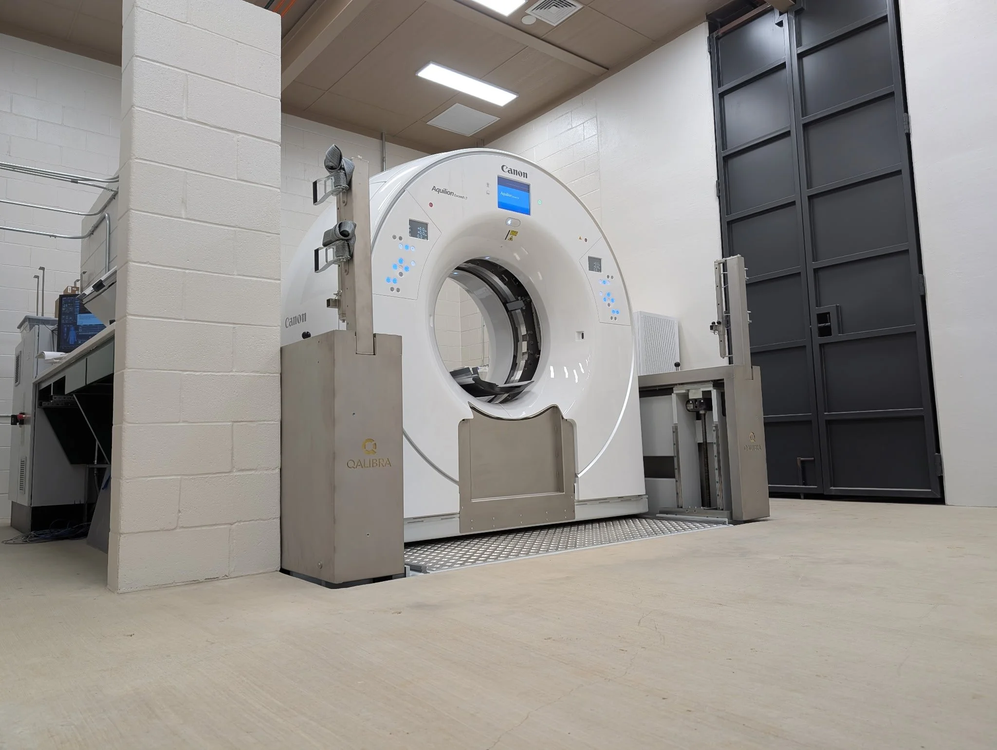
Computed Tomography (CT)
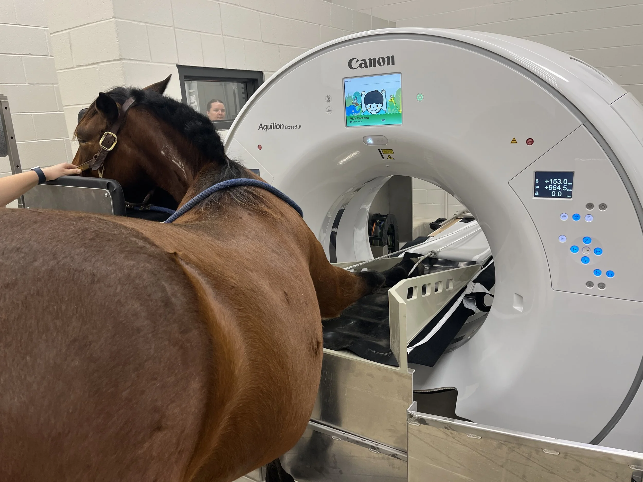
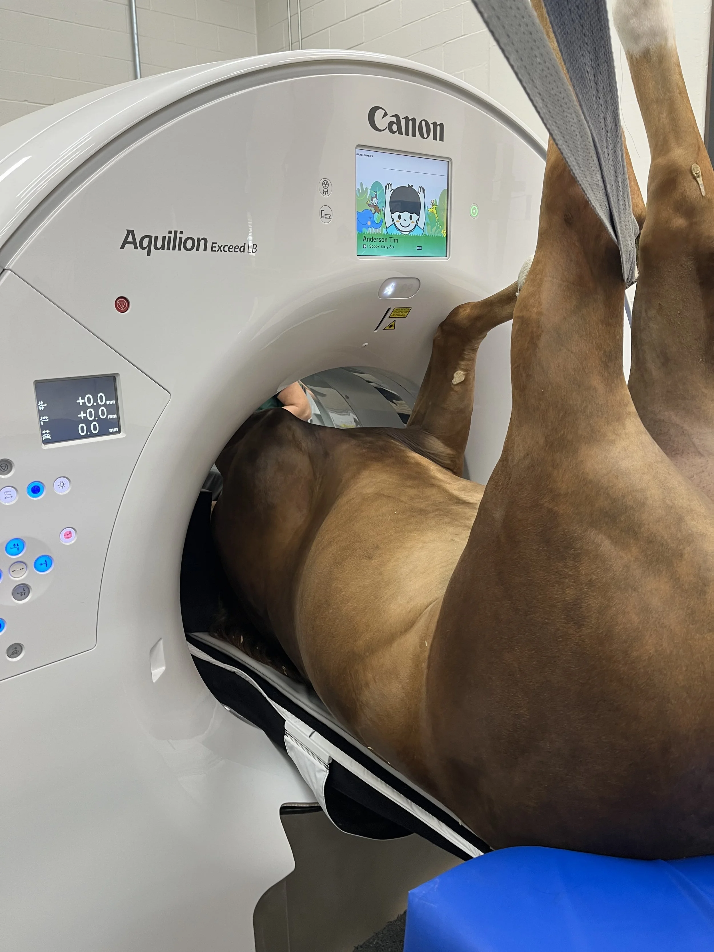
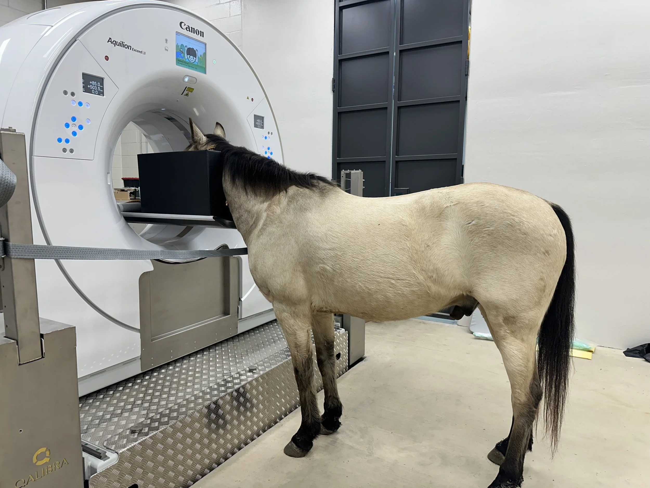
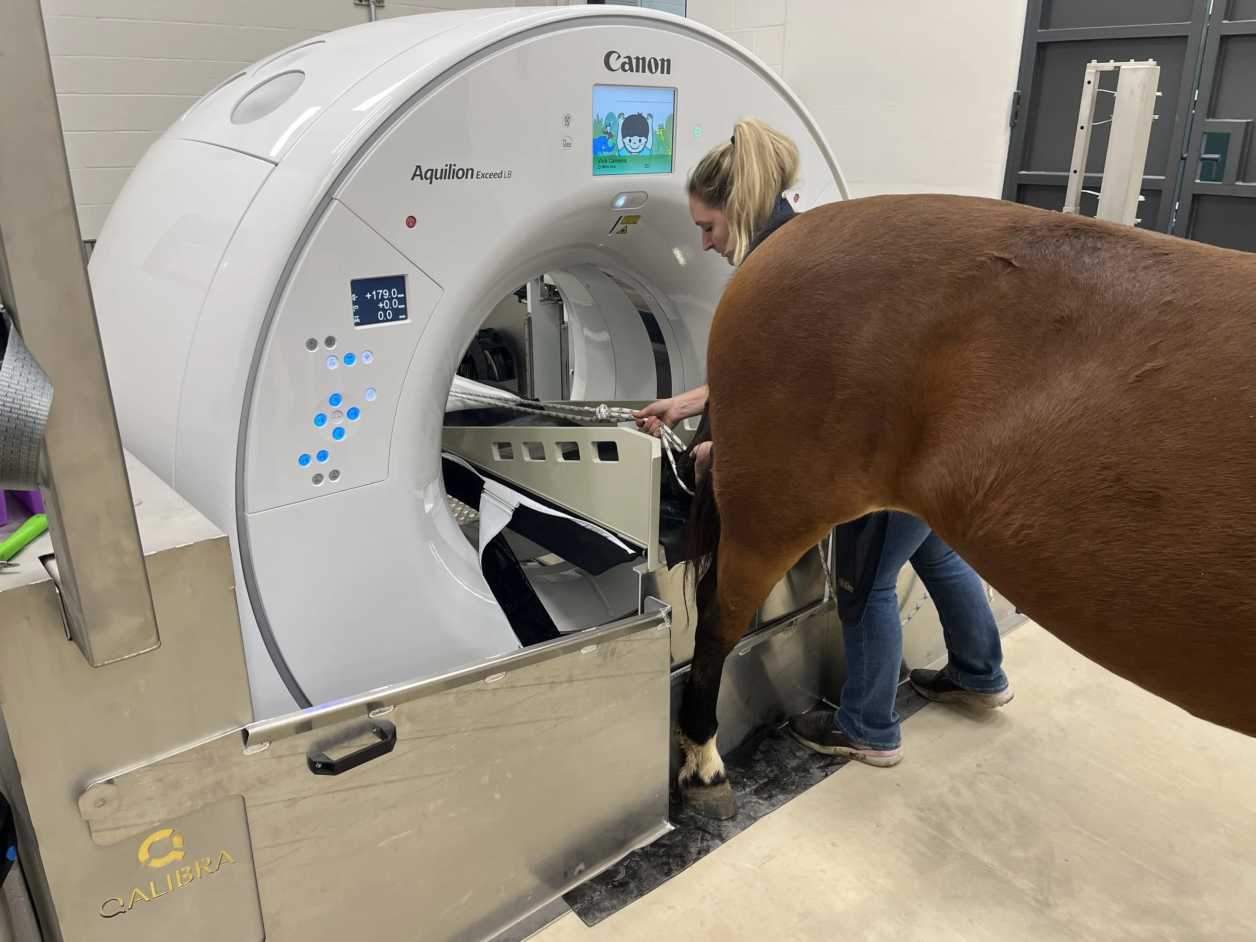
We are equipped with a Qalibra large bore CT machine made for the horse. This innovative design places the CT machine on a moving platform so that the machine moves around the horse. The height of the machine may also be lowered between ground level and the head height of an adult horse
Our CT machine is designed to allow for CT scans on horses both standing and under general anesthesia.
Standing exams are possible for heads, cranial cervical spine, and the distal limbs
General anesthesia is utilized to image the entire cervical spine including myelography, elbows, shoulders, stifles, and pelvis.